Brain Slice Transporter for the stable movement of viable slices between facilities

Description
The Brain Slice Transporter system was designed and developed to meet the needs of scientists working in laboratory areas that are physically separated from animal house facilities, or where live animals cannot be brought into the lab facility. In addition, there is an increasing demand for the transport of donated human tissue from clinical settings to be made available for laboratory experiments. Certain surgical procedures result in healthy tissue being discarded. Here, donated brain samples can be sectioned into thin slices and placed into the BST to maintain viability while being transported sometimes by road to a laboratory facility. If tissue samples are otherwise transported submerged, the motion of the aCSF causes movement and deterioration of the fragile sample. The BST addresses this by separating the moving fluid below from the tissue held in interface mode within a continuously perfused chamber whilst kept in high oxygen concentration and humidified in a closed container.
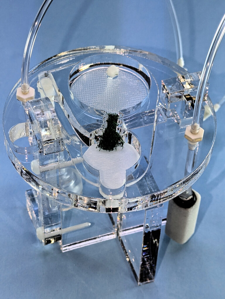
The transporter manifold is constructed in acrylic plastic, with an upper section containing an interface chamber, a solution feed reservoir and a liquid pump operated entirely by carbogen gas. The chamber is mounted above a reservoir containing oxygen saturated aCSF and contained in a transport jug with a handle for easy handling. A tight seal lid on top of the jug allows for the maintenance of a high oxygen and humidification level above the slices during transport. The diameter of the jug is 85mm, height is 160mm. The transporter module is carried entirely within the jug. The lower section of the jug maintains saturated aCSF with a volume of 500ml and provides humidification with an air stone incorporated into it. The aCSF is pumped up to the interface chamber above by means of a second carbogen line that feeds into a tube to allow bubbles to carry solution to the top, this is the fluid pump. This is fed to a reservoir from which aCSF is wicked into an interface chamber with a base lined with polypropylene mesh. Excess fluid returns from one end to the reservoir to the bottom, likewise extra solution from the reservoir returns to the bottom for re-circulation. Two separate carbogen feed tubes enter the jug from the lid and are connected to the upper chamber. An external source of portable carbogen is required for the operation of the Brain Slice Transporter system and the entire system is normally carried in a polystyrene ‘Igloo’ to maintain cool temperatures for the transport phase.
Features
- Designed for maintaining isolated, living slices in vitro
- Enables transportation of brain slices from surgical preparation areas to other laboratory locations
- Maintains oxygenation, circulation of artificial cerebrospinal fluid and humidification during transport
- Specifically addresses the need for maintaining viability during transport, even over distances involving walking or vehicular transportation
- Allows brain slices to remain in interface mode, minimizing disturbance from external motion
- Ensures slices remain viable throughout the transportation process
Schematic
Parts description
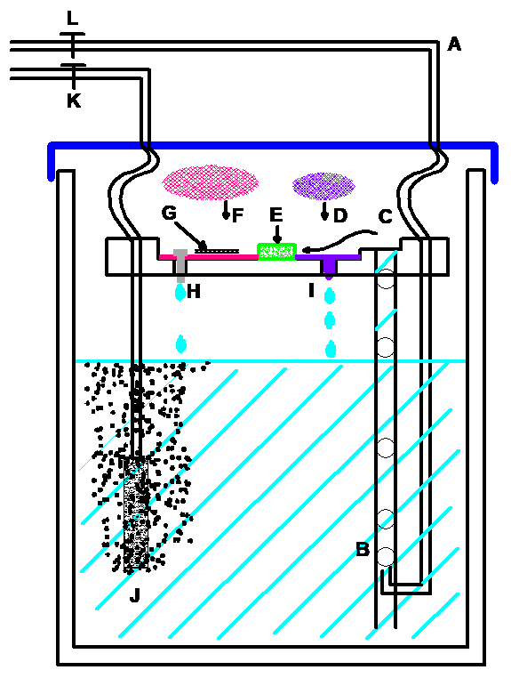
Carbogen enters the jar through the top of the lid at [A]
where it is connected to the bottom of a larger bore tube. Bubbles at [B]
rise up the tube taking aCSF to the top chamber part at [C].
A small polypropylene mesh disc [D]
with a drip control wick [I]
receives aCSF and is led to a buffer material [E].
A continuous feed of aCSF passes into another larger polypropylene mesh disc [F]
upon which the brain slice is laid over a piece of lens tissue [G].
Since the polypropylene mesh is wetted at it’s surface, lens tissue on which brain slices rest are maintained at an interface with high oxygen concentration with the lid in place
Excess aCSF falls through a drip point [H]
A second carbogen feed goes directly to an air stone bubbler [J]
Parts description
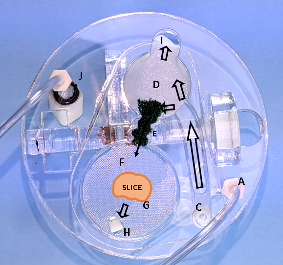
Carbogen enters the jar through the top of the lid at [A]
where it is connected to the bottom of a larger bore tube. Bubbles rise up the tube taking aCSF to the top chamber part at [C].
A small polypropylene mesh disc [D]
with a drip control wick where the mesh in bent down [I]
receives aCSF and is led to a buffer material [E].
A continuous feed of aCSF passes into another larger polypropylene mesh disc [F]
upon which the brain slice is laid over a piece of lens tissue [G].
Since the polypropylene mesh is wetted at its surface, the lens tissue on which brain slices rest are maintained at an interface with high oxygen concentration with the lid in place.
Excess aCSF falls through a drip point [H]
A second carbogen feed goes directly to an air stone bubbler [J]
to maintain a high concentration of oxygen.