Home > Ultrasound Imaging System

VINNO 6 LAB

The world’s first portable small animal ultrasound imaging system

Description

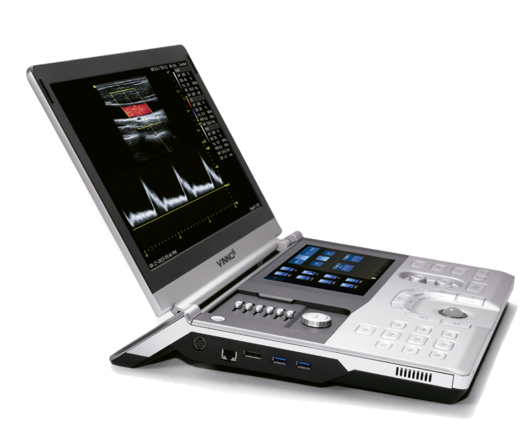
The small animal ultrasound imaging system is a comprehensive imaging platform. Compared with other imaging systems, the ultrasound system has the advantages of lower establishment, low maintenance costs, easy and fast operation, high efficiency and high-resolution imaging.

At the same time, the ultrasound system can provide anatomical information, functional imaging information and molecular imaging information, and can conduct long-term observation of the same animal, making the ultrasound system likely to become the most important auxiliary tool in small animal preclinical trials in the future.

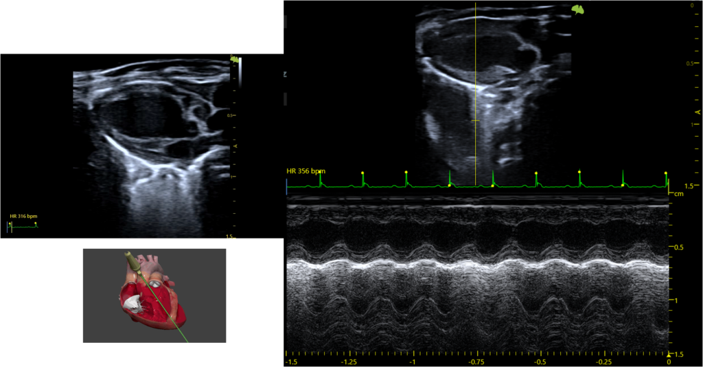
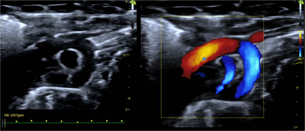
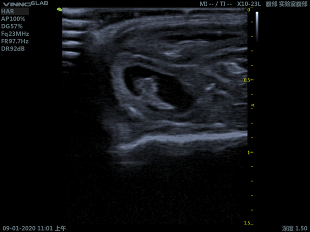
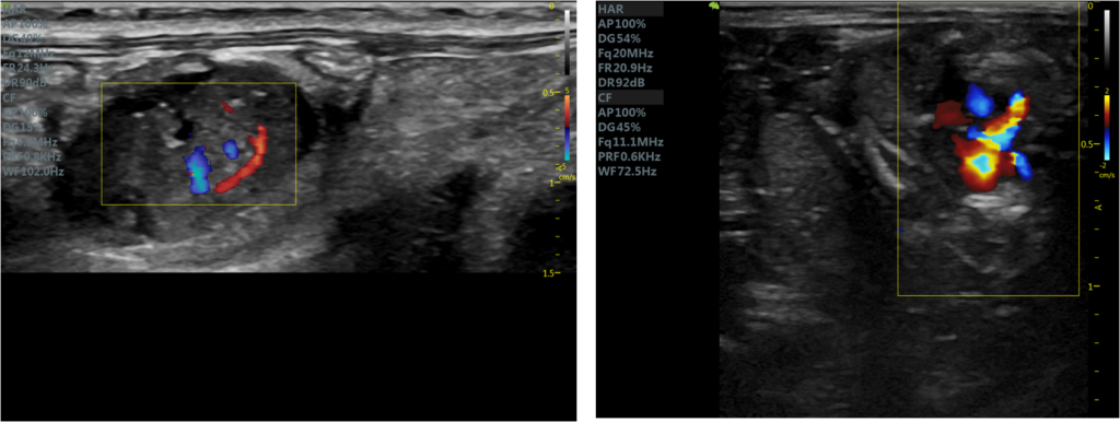
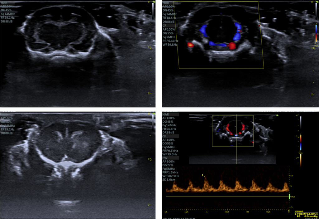
Ultrasound systems have been widely used in cardiovascular research (with the most comprehensive hemodynamic parameter measurement, cardiac structure and other functions), and are currently recognized as the gold standard in cardiovascular research. In addition, ultrasound systems have performed more prominently in many research areas such as oncology, developmental, musculoskeletal and metabolic diseases, as well as in embryology research. More than 2,000 articles have been published in peer-reviewed journals around the world (science, nature, circulation, cancer research, etc.). Today, ultrasound technology has been widely used in the human body clinically, which shows that it has more advantages in translational medicine.

It represents the development trend of new small animal molecular imaging, is the most promising diagnostic technology for clinical application, and is one of the indispensable technologies and tools in the fields of tumor, heart, blood vessel, and nerve research.

Features

  • The world’s first RF data transmission platform can collect raw diagnostic data 40 times higher than traditional technology, greatly improving image contrast and resolution

  • It has complete functions and can meet the needs of high-resolution imaging of the heart, blood vessels, brain, and abdominal organs and the measurement of various parameters. Most of the application functions are free and open to use

  • Covering a wide range of animal types, one machine can meet the application needs of all types of experimental animals, from mice to dogs, pigs, etc

  • Portable with thinner, lighter and compact construction

  • Data packet operation mode lowers the usage threshold and improves data consistency

  • Ergonomic and compact design for intuitive workflow

  • User friendly interface for easy and fast operation

  • Low establishment and maintenance costs

Application Cases

Cardiovascular

  • Cardiac structure
  • Congenital tissue defect
  • Cardiac function evaluation
  • Vascular function evaluation
PLAX, B-Mode and M-Mode

Angiology

  • Cross-sectional images of blood vessels
  • Measurement of the thickness of the arterial wall and the size of the lumen
  • Measurement of the intima, media, adventitia of the blood vessel wall
Aortic arch long-axis section, color Doppler

Oncology

  • Tumor detection and morphology
  • Measurement of parameters without the need of markers
  • Oncological pharmacology
Mouse Abdominal Tumor

Developmental and Reproductive

  • Prenatal screening
  • Genetic diseases
  • Reproductive function
Evaluation of cardiac function in 15-day-old fetal rats

Neuroscience

  • Brain tissues
  • Brain blood flow
  • Brain tumors
Mouse Brain Blood Flow

Gastrointestinal

  • High-resolution two-dimensional imagings
  • Real-time in vivo detection of blood flow and perfusion imaging
  • Liver, kidney, intestine and other tissues and organs of various animals such as rats and mice
  • Prenatal screening
  • Genetic diseases
  • Reproductive function
Mouse Kidney (left) and Rat Liver (right)

Downloads

currently no downloads available 

Get Support or A Quote

For immediate help on this product, please call: 1-289-4893376 

Otherwise, please use this web form 

On This Page